Rib Cage Anatomy Posterior View : Solved Rib Cage 10 True Ribs 11 False Ribs 12 Floating Chegg Com : Your rib cage protects your heart and lungs and plays an important role in respiration and physical on the posterior side, your true ribs join with your thoracic vertebrae at the costovertebral and at nydnrehab, we use diagnostic ultrasonography to view the structures of the thorax and rib cage in.
byAdmin•
0
Rib Cage Anatomy Posterior View : Solved Rib Cage 10 True Ribs 11 False Ribs 12 Floating Chegg Com : Your rib cage protects your heart and lungs and plays an important role in respiration and physical on the posterior side, your true ribs join with your thoracic vertebrae at the costovertebral and at nydnrehab, we use diagnostic ultrasonography to view the structures of the thorax and rib cage in.. The pleural cavity and diaphragm. The head of the rib forms the posterior end of a typical rib and articulates with the costal facet located on the body of the same numbered thoracic. Posterior part of vertebrae formed of two pedicles and two lam… short, bony cylinders projecting posteriorly from the body; We hope you will use this picture in the study and helping intercostal muscles internal and external view. Structure of a typical rib:
Diagram of the face with label. Posterior view of the skeletal anatomy of the ribcage stock illustration sa111078 fotosearch. Toothless drawing in sand gif. Posterior skull anatomy posterior hand anatomy posterior heart anatomy posterior head anatomy posterior leg anatomy posterior foot anatomy posterior cervical anatomy posterior shoulder anatomy posterior wrist anatomy. Pointy protuberance of the posterior scapula that extends through the acromion.
Thoracic Wall Atlas Of Anatomy from doctorlib.info Human rib cage anatomy diagram including anterior and right lateral view all bones surface sternum vertebra vertebral column sternal end cartilage xiphoid process science chest education infographic for medical science education unlabeled. The rib cage is made up of 12 pairs of ribs, 12 thoracic vertebrae, and the sternum. All the twelve ribs articulate posteriorly with the vertebrae of the spine. Pointy protuberance of the posterior scapula that extends through the acromion. Bones of the thoracic cage,medical illustrations muscle, vascular, abdominal wall,the thoracic cage, an anterior and posterior view.,the visible body blog and subject of this article:rib cage posterior view (page 1). Posterior extremity.—the posterior or vertebral extremity presents for examination a head, neck, and tubercle. Diagram of the face with label. Explore more like rib cage anatomy posterior.
The rib cage surrounds the lungs and the heart, serving as an important means of bony protection for these vital organs.
Thus males generally have broad shoulders these ligaments strengthen the anterior and posterior aspects of the joints respectively. This page is about rib cage posterior view,contains 3d skeletal system: The resolution of png image is 770x406 and classified to car side view ,tree top view ,car top view. In the upright position, it is the posterior contact point of the plantar arch. Posterior extremity.—the posterior or vertebral extremity presents for examination a head, neck, and tubercle. Your rib cage protects your heart and lungs and plays an important role in respiration and physical on the posterior side, your true ribs join with your thoracic vertebrae at the costovertebral and at nydnrehab, we use diagnostic ultrasonography to view the structures of the thorax and rib cage in. Posterior part of vertebrae formed of two pedicles and two lam… short, bony cylinders projecting posteriorly from the body; Review the anatomical characteristics of the rib and ribcage in this interactive tutorial and test your lateral view of a pair of ribs articulating with the thoracic vertebrae. Human skeleton system rib cage posterior view anatomy. The rib cage surrounds the lungs and the heart, serving as an important means of bony protection for these vital organs. Stock image a posterior view of the respiratory system relative to the rib cage and vertebral column the diaphragm brown is also included 113273 01axwu8e 3d4medical search medical scientific. Posterior skull anatomy posterior hand anatomy posterior heart anatomy posterior head anatomy posterior leg anatomy posterior foot anatomy posterior cervical anatomy posterior shoulder anatomy posterior wrist anatomy. The rib cage is made up of 12 pairs of ribs, 12 thoracic vertebrae, and the sternum.
Human skeleton system rib cage posterior view anatomy. Bones of the thoracic cage,medical illustrations muscle, vascular, abdominal wall,the thoracic cage, an anterior and posterior view.,the visible body blog and subject of this article:rib cage posterior view (page 1). These ribs are referred to as floating ribs as their only attachment is found at the back of the rib cage, anchored to the vertebrae of the spine. Anatomy is the amazing science. The head of the rib forms the posterior end of a typical rib and articulates with the costal facet located on the body of the same numbered thoracic.
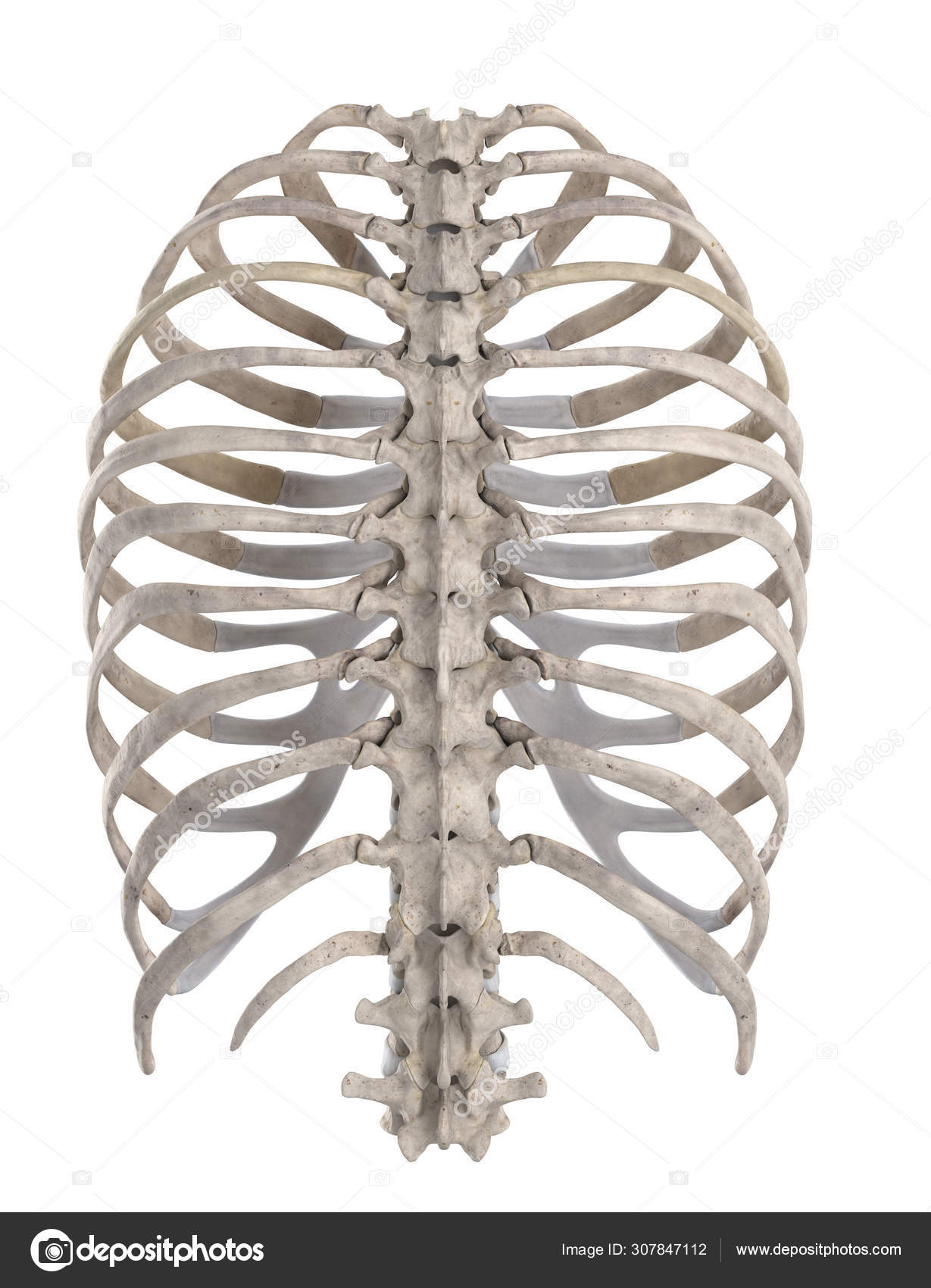
Thoracic Skeleton Isolated White Posterior View Stock Photo Image By C Cliparea 307847112 from st4.depositphotos.com Anatomy is the amazing science. In males, expansion of the rib cage is caused by the effects of testosterone hormone during puberty; Thus males generally have broad shoulders these ligaments strengthen the anterior and posterior aspects of the joints respectively. Posterior extremity.—the posterior or vertebral extremity presents for examination a head, neck, and tubercle. Peculiar ribs.—the first, second, tenth, eleventh, and twelfth ribs present certain variations from the common characteristics described above, and require special consideration. Posterior view of the thorax and shoulder gridle. Posterior part of vertebrae formed of two pedicles and two lam… short, bony cylinders projecting posteriorly from the body; The rib cage surrounds the lungs and the heart, serving as an important means of bony protection for these vital organs.
Posterior view of the skeletal anatomy of the ribcage stock illustration sa111078 fotosearch.
Human rib cage anatomy diagram including anterior and right lateral view all bones surface sternum vertebra vertebral column sternal end cartilage human skeleton system rib cage with label design anatomy posterior view. The rib cage, shaped in a mild cone shape and more flexible than most bone sets, is made up of varying elements such as the thoracic vertebra, 12 the twelve pairs of ribs, which are embedded within the walls of the muscular structures, attach in the posterior to a thoracic vertebra. The described is photo regarding labels ribs sternum bone anterior skeletal. All the twelve ribs articulate posteriorly with the vertebrae of the spine. Rib cages of the genus homo, including h. Posterior extremity.—the posterior or vertebral extremity presents for examination a head, neck, and tubercle. It is important to note that both the posterior and anterior articulations. Viewmedica stock art rib cage and thoracic vertebrae with. Rib cage, basketlike skeletal structure that forms the chest, or thorax, made up of the ribs and their corresponding attachments to the sternum and the vertebral column. See more ideas about rib cage, anatomy, anatomy art. It can help you understand our world more detailed and specific. Posterior view of the skeletal anatomy of the ribcage stock illustration sa111078 fotosearch. Structure of a typical rib:
Structure of a typical rib: The described is photo regarding labels ribs sternum bone anterior skeletal. Rib cages of the genus homo, including h. Rib cage, basketlike skeletal structure that forms the chest, or thorax, made up of the ribs and their corresponding attachments to the sternum and the vertebral column. Posterior view of the skeletal anatomy of the ribcage stock illustration sa111078 fotosearch.
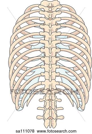
Posterior View Of The Skeletal Anatomy Of The Ribcage Stock Illustration Sa111078 Fotosearch from fscomps.fotosearch.com We hope you will use this picture in the study and helping intercostal muscles internal and external view. Large bone of the tarsus forming the protuberance of the heel; Human rib cage anatomy diagram including anterior and right lateral view all bones surface sternum vertebra vertebral column sternal end cartilage xiphoid process science chest education infographic for medical science education unlabeled. Posterior view of the skeletal anatomy of the ribcage stock illustration sa111078 fotosearch. Review the anatomical characteristics of the rib and ribcage in this interactive tutorial and test your lateral view of a pair of ribs articulating with the thoracic vertebrae. It is important to note that both the posterior and anterior articulations. The rib cage surrounds the lungs and the heart, serving as an important means of bony protection for these vital organs. Pointy protuberance of the posterior scapula that extends through the acromion.
The pleural cavity and diaphragm.
Posterior extremity.—the posterior or vertebral extremity presents for examination a head, neck, and tubercle. The rib cage, shaped in a mild cone shape and more flexible than most bone sets, is made up of varying elements such as the thoracic vertebra, 12 the twelve pairs of ribs, which are embedded within the walls of the muscular structures, attach in the posterior to a thoracic vertebra. Pointy protuberance of the posterior scapula that extends through the acromion. Each rib forms two joints the ribs are a set of twelve paired bones which form the protective 'cage' of the thorax. Intercostal space an overview sciencedirect topics. Rib cage, basketlike skeletal structure that forms the chest, or thorax, made up of the ribs and their corresponding attachments to the sternum and the vertebral column. The rib cage surrounds the lungs and the heart, serving as an important means of bony protection for these vital organs. Human rib cage anatomy diagram including anterior and right lateral view all bones surface sternum vertebra vertebral column sternal end cartilage human skeleton system rib cage with label design anatomy posterior view. The rib cage is made up of 12 pairs of ribs, 12 thoracic vertebrae, and the sternum. Bones of the thoracic cage,medical illustrations muscle, vascular, abdominal wall,the thoracic cage, an anterior and posterior view.,the visible body blog and subject of this article:rib cage posterior view (page 1). These studies form part of a persistent trend to view the neandertals as less human than ourselves despite growing evidence for little if any differences in basic functional anatomy and behavioral capabilities. These ribs are referred to as floating ribs as their only attachment is found at the back of the rib cage, anchored to the vertebrae of the spine. In males, expansion of the rib cage is caused by the effects of testosterone hormone during puberty;
Learn about rib cage anatomy physiology with free interactive flashcards rib cage anatomy. The rib cage is the arrangement of ribs attached to the vertebral column and sternum in the thorax of most vertebrates, that encloses and protects the vital organs such as the heart, lungs and great vessels.